
Machine Learning in Medicine
Using AI to diagnose endometriosis without surgery

Generative artificial intelligence (AI) technologies such as ChatGPT have a huge potential to disrupt traditional methods of communication. AI technology can also be used to improve the diagnosis, treatment and monitoring of serious medical conditions in patients.
One of these is endometriosis, which affects an estimated one in nine women worldwide.
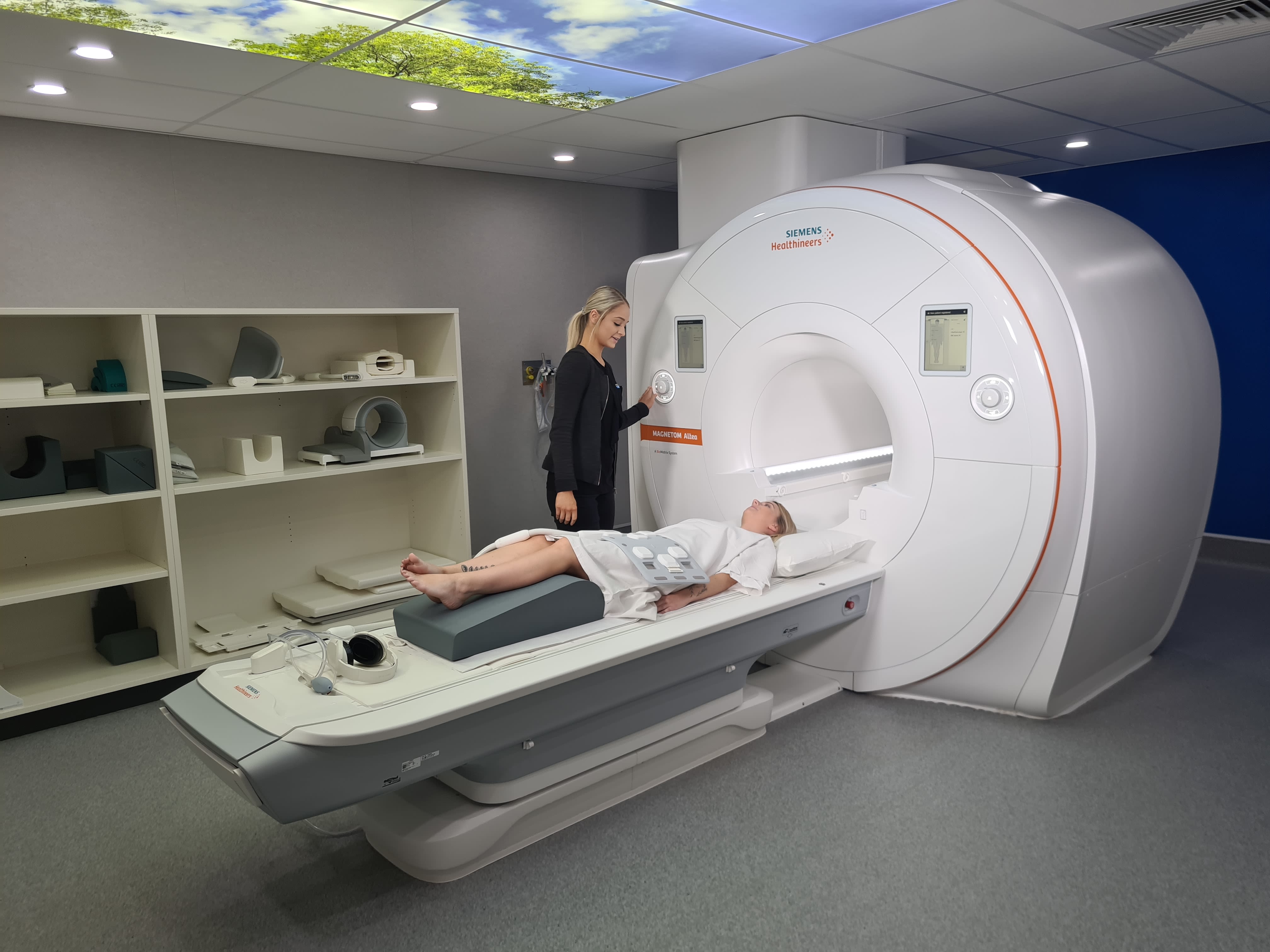
Two University of Adelaide research institutes – the Robinson Research Institute and the Australian Institute for Machine Learning – are working together to harness artificial intelligence to help change the way endometriosis is diagnosed. By combining ultrasound, magnetic resonance imaging (MRI) and AI, the new technology aims to provide a cost-effective, accessible, and accurate way to diagnose the condition without surgery.

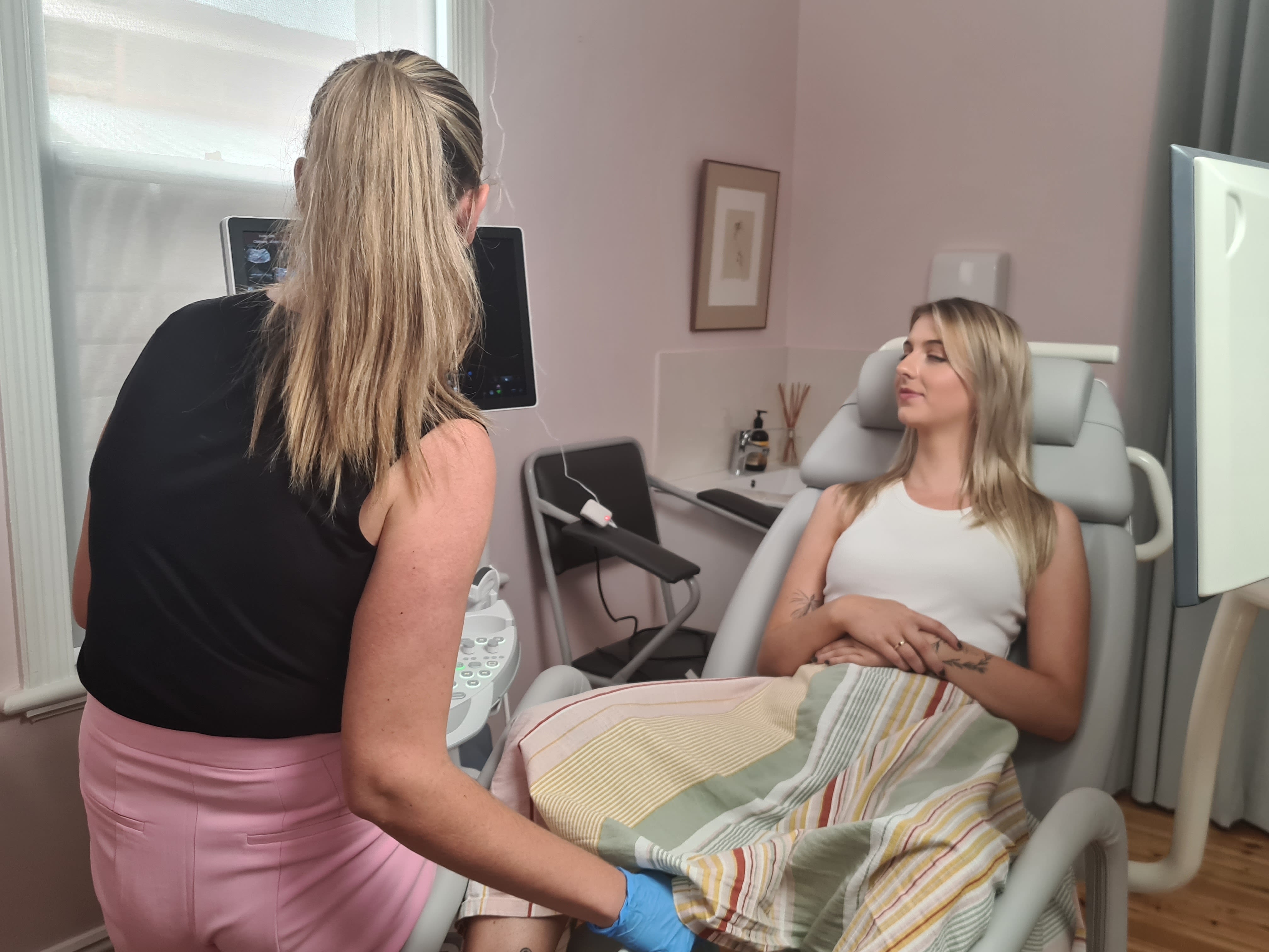
Specialist Gynaecological sonographer Alison Deslandes preparing for a transvaginal ultrasound on endometriosis patient Belle
Specialist Gynaecological sonographer Alison Deslandes preparing for a transvaginal ultrasound on endometriosis patient Belle
The endometrium is the layer of spongy tissue that lines the uterus. Endometriosis is a condition where cells similar to this lining grow outside the uterus causing severe pain and fertility problems. Diagnosis is often delayed, with several years between the onset of symptoms and diagnosis.
Currently, endometriosis is usually diagnosed by keyhole surgery – which is costly, painful and time consuming. Surgery is difficult to access and many young people find their symptoms are dismissed and treatment is delayed and unfocused without an endometriosis diagnosis. Professor Louise Hull, a gynaecologist and fertility specialist at the Robinson Research Institute, saw an opportunity to develop less invasive methods. “It’s something people have really needed and wanted for a very long time,” she says.
Specialist scans, such as transvaginal ultrasound and gynaecological MRIs, have the potential to identify diagnostic markers that indicate endometriosis. The challenge, however, comes in interpreting the images. “Endometriosis scanning is a specialist skill and not all sonographers, radiologists and gynaecologists will have the opportunity to receive training to perform specialist endometriosis scans or MRIs,” says Professor Hull.
The Adelaide research team received an AUD $2 million grant from the Australian Government’s Medical Research Future Fund in July 2021. The resulting study used machine learning to automatically combine the digital diagnostic capabilities of pelvic scans and magnetic resonance imaging in order to identify endometriosis lesions.
Using a diagnostic dataset provided by Professor George Condous as a training sample, researchers at the Australian Institute for Machine Learning built a program that can read specialist scans and recognise certain endometriosis imaging markers, helping doctors to provide a diagnosis. The outcome is IMAGENDO® – an AI-generated diagnostic algorithm that accurately estimates the likelihood that an individual has endometriosis based on specialist ultrasound or MRI scans.
Initial tests show that the software is capable of diagnostic accuracy approaching that of a specialist endometriosis doctor, which will improve as the research develops. “Machine learning is an iterative process – as you add more and more training samples, the accuracy of the system improves,” says Professor Gustavo Carneiro.
The Robinson Research Institute has collaborated with the Australian Institute of Machine Learning to use large numbers of ultrasounds and MRIs and put all the digital data together. “A non-invasive imaging diagnosis means people can explore different treatment options for best care, including non- invasive options such as physiotherapy and medications as well as surgery,” says Professor Hull.
“Surgery is still a critical aspect of endometriosis care but having an accurate assessment of where endometriosis lesions are located can also improve the quality of surgery. It is easier for surgeons to effectively treat people in one operation if they know where the lesions are, and they can ensure the right equipment and doctors (such as bowel surgeons) are present. This avoids repeat surgeries and prolonged surgical treatments for people with endometriosis.”

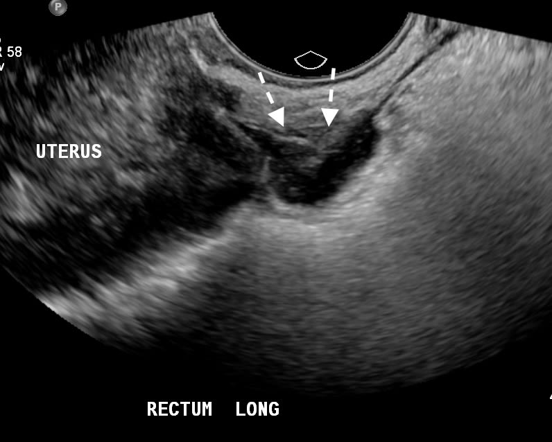

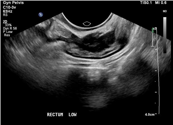

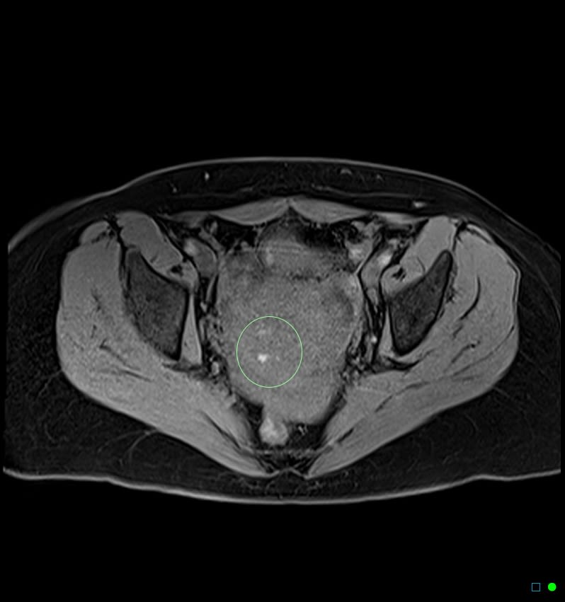

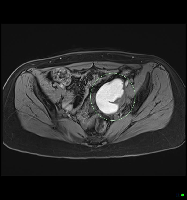
Ultrasound and MRI scans showing endometriosis lesions


The IMAGENDO® team including Professor Louise Hull (front row, second from left) and Dr Jodie Avery (front row, second from right)
The IMAGENDO® team including Professor Louise Hull (front row, second from left) and Dr Jodie Avery (front row, second from right)
The IMAGENDO® technology is also timely – the European Society of Human Reproduction and Embryology, the National Institute for Health and Care Excellence and the Royal Australian and New Zealand College of Obstetricians and Gynaecologists have all recently agreed that specialist ultrasound and MRI scans can be used to diagnose endometriosis quite accurately, although a negative ultrasound or MRI can never exclude endometriosis. The team is exploring different ways that the model could be rolled out once the algorithm has been tested and approved for use, with an aim of it being available in clinics around the world in the next 5-10 years.
Program manager Dr Jodie Avery can see clear benefits of the technology. “Endometriosis symptoms can have a devastating impact on women’s lives.”
“We hope that an earlier diagnosis enabled by the IMAGENDO® technology will lead to prompt treatment and better quality of life by avoiding unnecessary hospitalizations and repetitive surgery.”
“It provides an opportunity to manage and treat pain appropriately to prevent complications and to preserve fertility via egg freezing and fertility planning. Having a reliable diagnosis also helps the community to provide appropriate support for people with endometriosis. This improves mental health, engagement with education and work, and the ability to participate in everyday life.”
IMAGENDO® is increasingly recognized nationally and internationally for the way it has used AI to integrate digital data from ultrasound and MRI scans to formulate new diagnostics for endometriosis. In August, the team were awarded a prestigious Australian Museum Eureka Prize – the country’s most comprehensive national science awards – for Innovative Use of Technology. The program is expanding nationally in Australia and internationally with collaborations in North America, South America and in Europe.
About the University of Adelaide:

This content was paid for and created by The University of Adelaide. The editorial staff at The Chronicle had no role in its preparation. Find out more about paid content.